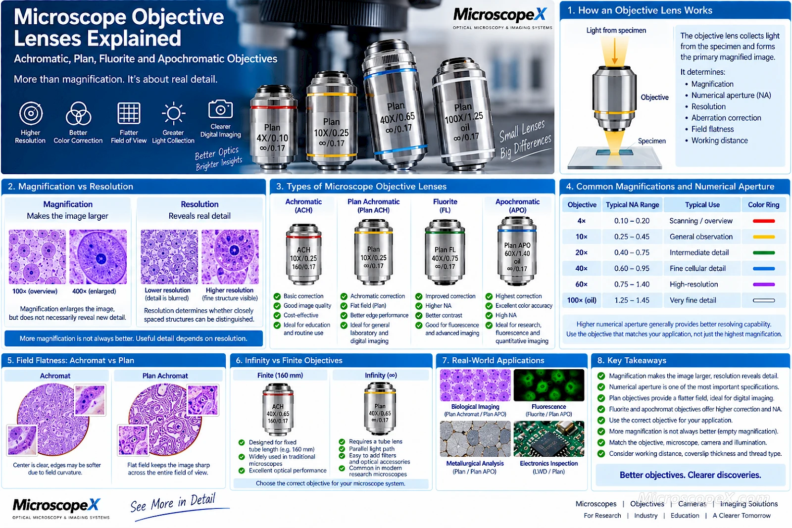
The objective lens is one of the most important components of an optical microscope.
It determines much more than magnification.
A microscope objective strongly influences:
- Optical resolution
- Numerical aperture
- Image contrast
- Color correction
- Field flatness
- Working distance
- Light-gathering capability
- Imaging performance
- Camera image quality
This is why two objectives labeled 40× can produce noticeably different images.
One may be a basic achromatic objective.
Another may be a plan achromatic objective.
A higher-performance system might use a fluorite or plan fluorite objective.
At the upper end, a plan apochromatic objective can provide significantly improved correction, higher numerical aperture and excellent performance for demanding digital imaging.
Understanding these terms makes it much easier to select the correct objective for biological microscopy, metallurgical inspection, fluorescence imaging, digital microscopy and research.
Explore the current Microscope Objectives available from MicroscopeX.
What Does a Microscope Objective Do?
The objective is the lens assembly positioned closest to the specimen.
It collects light from the specimen and creates the primary magnified optical image.
In a compound microscope, the objective is responsible for a large part of the system's:
- Magnification
- Resolution
- Numerical aperture
- Aberration correction
The eyepiece then enlarges the image created by the objective.
This is an important distinction.
A higher-power eyepiece can make an image larger, but it cannot compensate for poor objective resolution.
For this reason:
The objective is usually more important to image quality than the eyepiece.
Common Microscope Objective Magnifications
Biological microscopes commonly use objectives such as:
- 4×
- 10×
- 20×
- 40×
- 60×
- 100×
Metallurgical and industrial microscope systems may also use:
- 2×
- 5×
- 10×
- 20×
- 50×
- 80×
- 100×
Each magnification provides a different balance between:
- Field of view
- Resolution
- Working distance
- Depth of field
A microscope should therefore use the objective appropriate for the feature being observed rather than simply the highest available magnification.
How Total Microscope Magnification Is Calculated
For a conventional compound microscope:
Total Magnification = Objective Magnification × Eyepiece Magnification
For example:
| Objective | Eyepiece | Total Magnification |
|---|---|---|
| 4× | 10× | 40× |
| 10× | 10× | 100× |
| 20× | 10× | 200× |
| 40× | 10× | 400× |
| 100× | 10× | 1000× |
However, objective performance cannot be judged by magnification alone.
The next specification you should examine is numerical aperture.
What Is Numerical Aperture?
Numerical aperture is abbreviated as:
NA
It describes an objective's ability to collect light and resolve fine detail.
In simplified form:
NA = n × sin(θ)
where:
- n is the refractive index of the medium between specimen and objective
- θ is related to the angle of light accepted by the objective
Higher numerical aperture generally provides:
- Better resolving capability
- Greater light collection
- Finer specimen detail
This is why two 40× objectives can perform differently.
For example:
40× / 0.65
and:
40× / 0.95
have the same magnification but very different numerical apertures.
The 0.95 NA objective can generally resolve finer structures.
Magnification Does Not Equal Resolution
A common mistake is to assume:
Higher magnification = higher resolution
This is not always true.
Magnification determines how large the image appears.
Resolution determines whether two closely spaced specimen features can actually be distinguished.
A high-quality 40× objective with a high NA may reveal more useful detail than a lower-quality objective combined with excessive eyepiece or digital magnification.
For a deeper explanation, see:
Microscope Magnification vs Resolution
Understanding Objective Correction Classes
Microscope objectives can be divided into several broad optical correction classes.
The most common terms are:
- Achromatic
- Plan Achromatic
- Fluorite
- Plan Fluorite
- Apochromatic
- Plan Apochromatic
These terms describe how well the objective corrects optical aberrations.
In general, optical correction increases approximately in this direction:
Achromat
↓
Plan Achromat
↓
Fluorite / Plan Fluorite
↓
Apochromat / Plan Apochromat
Higher correction usually means:
- Better color correction
- Better spherical correction
- Higher potential NA
- Better edge performance when plan-corrected
- Higher cost
- More complex optical design
What Is Chromatic Aberration?
Different wavelengths of light do not refract by exactly the same amount when passing through glass.
Without correction, red, green and blue light may focus at slightly different positions.
This can produce:
- Colored fringes
- Reduced sharpness
- Lower contrast
- Inaccurate color reproduction
This optical defect is called chromatic aberration.
Objective designers use combinations of different glass types and lens elements to bring multiple wavelengths into better focus.
The degree of correction varies by objective class.
What Is Spherical Aberration?
A simple spherical lens does not focus all incoming rays at exactly the same point.
Light passing through different parts of the lens can focus at slightly different axial positions.
This is called spherical aberration.
It can reduce:
- Sharpness
- Contrast
- Fine-detail reproduction
More sophisticated objective designs include additional lens groups to control spherical aberration.
Higher correction classes generally provide better spherical correction.
What Is Field Curvature?
A microscope specimen is usually flat.
But an optical system can naturally produce an image whose best-focus surface is curved.
This creates a practical problem:
Center in focus
Edges may be slightly out of focus.
Or:
Edges in focus
Center may no longer be perfectly focused.
This is called field curvature.
Plan objectives are specifically designed to improve this problem.
What Does “Plan” Mean?
The word:
Plan
indicates improved flat-field correction.
A plan objective is designed so that a much larger portion of the visible image field remains in focus simultaneously.
This is particularly important for:
- Microscope cameras
- Digital imaging
- Photomicrography
- Measurement
- Whole-field documentation
- Large camera sensors
A non-plan objective may still produce an excellent image near the center.
But edge focus may be weaker.
Achromat vs Plan Achromat
This is one of the most important objective comparisons for everyday microscopy.
Achromatic Objective
A basic achromatic objective provides fundamental correction for chromatic and spherical aberrations.
Advantages include:
- Affordable
- Simple
- Reliable
- Good central image quality
- Suitable for routine observation
Typical applications:
- Education
- Basic biological microscopy
- Routine laboratory work
- General observation
MicroscopeX offers traditional biological achromatic objective configurations such as the 195 mm Achromatic Biological Objectives.
Limitations of Achromatic Objectives
Compared with more highly corrected objectives, a basic achromat may show:
- Less flatness toward image edges
- More residual chromatic aberration
- Lower numerical aperture at some magnifications
- Lower performance for large-sensor digital imaging
This does not make achromatic objectives “bad.”
They are often entirely sufficient for routine visual microscopy.
The question is whether the application requires more correction.
What Is a Plan Achromatic Objective?
A plan achromatic objective combines:
Achromatic correction
with:
Flat-field correction
This means it provides a flatter usable image across the microscope field.
Advantages include:
- Better edge focus
- Better digital imaging
- Improved whole-field observation
- Good balance of performance and cost
Plan achromats are now extremely common in professional biological and industrial microscopy.
Achromat vs Plan Achromat
| Feature | Achromat | Plan Achromat |
|---|---|---|
| Chromatic correction | Good | Good |
| Spherical correction | Basic | Basic to improved |
| Field flatness | Limited | Significantly improved |
| Center image | Good | Good |
| Edge image | Less consistent | Better |
| Digital imaging | Basic | Very good |
| Cost | Lower | Moderate |
| Routine observation | Excellent | Excellent |
| Photography | Usable | Better |
For most modern digital microscope systems, plan achromatic objectives provide an excellent balance between cost and image quality.
Why Plan Objectives Matter More with Cameras
When looking through eyepieces, your eyes naturally concentrate on the center of the field.
You may not notice mild edge defocus.
A camera records the entire rectangular sensor simultaneously.
Therefore, field curvature becomes more obvious.
This is especially important when using:
- Large sensors
- High-resolution cameras
- Image stitching
- Measurements
- Digital documentation
For camera-based microscopy, a plan objective can provide a visibly more consistent image.
MicroscopeX Plan Achromatic Biological Objectives
For biological microscopy, MicroscopeX offers Infinity Plan Achromatic Biological Objectives.
The current series includes:
- 4× / NA 0.10
- 10× / NA 0.25
- 20× / NA 0.40
- 40× / NA 0.65
- 60× / NA 0.80
- 100× / NA 1.25 oil
This range covers routine scanning through high-resolution oil-immersion observation.
What Is a Fluorite Objective?
Fluorite objectives occupy a performance level between achromatic and apochromatic objectives.
They may also be described as:
Semi-apochromatic objectives
or by manufacturer-specific terms such as:
Fluor
or:
Fluar
Historically, fluorite-containing optical materials were used because of their favorable dispersion characteristics.
Modern high-performance objectives may use advanced optical glasses or synthetic materials to achieve similar or better correction.
Advantages of Fluorite Objectives
Compared with basic achromats, fluorite objectives generally provide:
- Better chromatic correction
- Better spherical correction
- Higher numerical aperture
- Better contrast
- Better resolution
- Improved light collection
These characteristics make fluorite objectives useful for:
- Fluorescence microscopy
- Digital imaging
- Research
- High-quality photomicrography
- Low-light applications
Why Fluorite Objectives Are Useful for Fluorescence
Fluorescence imaging often involves weak light.
Higher numerical aperture is valuable because it allows the objective to collect more emitted fluorescence.
A fluorite objective may therefore provide:
- Brighter fluorescence signals
- Better resolving capability
- Stronger contrast
than a basic achromat of the same magnification.
However, actual fluorescence performance also depends on:
- Optical transmission
- Coatings
- Autofluorescence characteristics
- Camera sensitivity
- Illumination
The objective should be selected as part of the complete fluorescence imaging system.
What Is a Plan Fluorite Objective?
A plan fluorite combines:
Fluorite-level optical correction
with:
Flat-field correction
The result is a strong middle-to-high-end objective class suitable for:
- Digital photography
- Fluorescence
- Research
- High-resolution biological imaging
Plan fluorites often provide an attractive balance between:
Performance
and:
Cost
when a full plan apochromat is unnecessary.
What Is an Apochromatic Objective?
An apochromatic objective represents a higher level of optical correction.
Traditionally, apochromatic designs bring at least three widely separated colors into substantially improved common focus while also providing advanced spherical correction.
Modern high-performance APO designs may correct even more wavelengths.
The practical result can include:
- Reduced color fringing
- Higher contrast
- Higher numerical aperture
- Better fine-detail reproduction
- Improved color accuracy
Apochromats are among the highest-performance objectives used in conventional optical microscopy.
Why Apochromatic Objectives Are Expensive
An apochromatic objective typically requires:
- More lens elements
- More specialized optical glass
- More complex correction
- Tighter manufacturing tolerances
- More precise alignment
The optical design may need to simultaneously control:
- Chromatic aberration
- Spherical aberration
- Coma
- Astigmatism
- Distortion
while maintaining high numerical aperture.
This complexity increases manufacturing cost.
What Is a Plan Apochromatic Objective?
A plan apochromat combines:
Advanced chromatic correction
Advanced spherical correction
Flat-field correction
This is one of the highest-performance general objective classes.
Advantages include:
- Excellent field flatness
- High numerical aperture
- Strong resolution
- Excellent color correction
- Excellent digital imaging
- Excellent photomicrography
These objectives are especially suitable for demanding applications.
Typical Applications for Plan Apochromats
Plan apochromatic objectives are commonly selected for:
- Research microscopy
- High-resolution digital imaging
- Fluorescence
- Materials science
- Semiconductor inspection
- Quantitative imaging
- High-quality photomicrography
They are particularly valuable when image quality must remain high across a large sensor field.
MicroscopeX Full APO Metallurgical Objectives
MicroscopeX offers Full APO Infinity Metallurgical Objectives designed for infinity-corrected metallurgical microscopy.
Available configurations include magnifications such as:
- 2×
- 5×
- 10×
- 20×
- 50×
- 100×
The series combines:
- Infinity correction
- Plan apochromatic design
- Long working distance
- Metallurgical imaging
This makes the objectives suitable for demanding industrial and materials applications.
Achromat vs Fluorite vs Apochromat
A simplified comparison looks like this:
| Feature | Achromat | Fluorite | Apochromat |
|---|---|---|---|
| Optical correction | Basic | Advanced | Highest |
| Chromatic correction | Good | Better | Excellent |
| Spherical correction | Basic | Better | Excellent |
| Typical NA | Lower | Medium-high | Often highest |
| Contrast | Good | Very good | Excellent |
| Resolution potential | Good | Very good | Excellent |
| Fluorescence | Basic | Very good | Excellent |
| Photography | Good | Very good | Excellent |
| Cost | Lowest | Medium-high | Highest |
Remember that:
Plan
is a separate flat-field correction designation.
Therefore:
- Plan Achromat
- Plan Fluorite
- Plan Apochromat
offer better field flatness than their non-plan equivalents.
A Practical Objective Quality Ladder
A simplified way to think about the categories is:
Achromat
Routine observation
↓
Plan Achromat
Routine professional imaging
↓
Plan Fluorite
Advanced imaging and fluorescence
↓
Plan Apochromat
High-end research and quantitative imaging
This is not an absolute ranking for every application.
A specialized long-working-distance plan achromat may be more appropriate than an APO objective for some industrial tasks.
The best objective is the one that matches the application.
What Is an Infinity-Corrected Objective?
Modern microscope objectives are frequently marked with:
∞
This indicates an infinity-corrected optical system.
In an infinity microscope, the objective sends light toward the tube lens as an approximately parallel beam.
The tube lens then forms the intermediate image.
The basic optical path is:
Specimen
↓
Infinity Objective
↓
Parallel Optical Space
↓
Tube Lens
↓
Intermediate Image
↓
Eyepiece / Camera
Why Infinity Correction Is Useful
Infinity optical systems provide several important advantages.
Optical components can be inserted into the parallel beam path with less disruption to focus.
Examples include:
- Beam splitters
- Fluorescence filters
- Polarizers
- DIC components
- Camera modules
- Measurement optics
This makes infinity systems highly modular.
They are common in modern:
- Research microscopes
- Fluorescence microscopes
- Metallurgical microscopes
- Industrial imaging systems
Infinity Objectives Require the Correct Tube Lens
An infinity objective does not independently form the final intermediate image at a fixed tube length.
It requires a compatible tube lens.
Therefore:
An infinity objective should not be assumed to work correctly on any microscope simply because the thread fits.
Compatibility depends on:
- Tube lens focal length
- Optical design
- Objective correction strategy
- Mechanical interface
Using an infinity objective on an incompatible microscope can produce incorrect magnification or poor optical correction.
What Is a Finite-Conjugate Objective?
Traditional microscopes often use finite tube-length objectives.
These may be marked with values such as:
160
or another tube-length specification.
In a finite-conjugate system, the objective forms the intermediate image directly at a specified distance.
A typical optical path is:
Specimen → Objective → Intermediate Image → Eyepiece
Finite systems can provide excellent performance.
They are simply designed around a different optical architecture.
Infinity vs Finite Objectives
| Feature | Finite Objective | Infinity Objective |
|---|---|---|
| Optical design | Fixed conjugate distance | Requires tube lens |
| Intermediate optical space | Limited | Parallel infinity space |
| Accessories | Less flexible | Highly flexible |
| Modern research systems | Less common | Very common |
| Cost | Often lower | Often higher |
| Compatibility | System-specific | System-specific |
One is not automatically “better.”
The objective must match the microscope system for which it was designed.
Never Mix Objective Systems Without Checking Compatibility
A microscope objective may physically screw into the nosepiece but still be optically incompatible.
Potential problems include:
- Incorrect magnification
- Poor parfocality
- Color errors
- Spherical aberration
- Reduced image quality
Always check:
- Objective thread
- Tube system
- Tube-lens design
- Parfocal distance
before changing objective families.
What Is RMS Objective Thread?
Many biological microscope objectives use the traditional RMS thread.
RMS is approximately:
20.32 mm diameter
with:
36 threads per inch
MicroscopeX biological objective families such as the Infinity Biological Objectives use the standard RMS-type 20.2 mm interface.
Metallurgical Objective Threads
Industrial and metallurgical objectives may use different mechanical interfaces.
For example, some infinity BF/DF metallurgical objectives use approximately:
26 mm threaded interfaces
rather than standard biological RMS.
Therefore:
Do not assume that every microscope objective uses the same thread.
Always confirm the mechanical specification.
Brightfield vs Brightfield/Darkfield Objectives
Metallurgical microscopes may use specialized objectives designed for:
BF
or:
BF/DF
meaning:
- Brightfield
- Brightfield / Darkfield
Darkfield reflected-light microscopy requires a different illumination geometry.
A BF/DF objective must accommodate the required illumination path.
MicroscopeX offers Infinity BF/DF Plan Achromatic Metallurgical Objectives for this type of system.
Why Metallurgical Objectives Are Different
Biological microscopes commonly observe transparent specimens using transmitted light.
Metallurgical microscopes often examine opaque objects using reflected illumination.
Typical samples include:
- Metals
- Semiconductor wafers
- Coatings
- Circuit structures
- Ceramics
- Polished materials
Metallurgical objectives may therefore be designed for:
- No cover glass
- Reflected-light imaging
- Longer working distance
- BF/DF illumination
- Industrial measurement
This is why a biological objective should not automatically be substituted for a metallurgical one.
What Is Working Distance?
Working distance is the distance between the front of the objective and the specimen when the specimen is in focus.
This specification becomes particularly important at higher magnifications.
As magnification and NA increase, working distance often decreases.
For example, an objective may have:
- Several millimeters of working distance at low magnification
- Less than 1 mm at high magnification
This affects usability.
Why Working Distance Matters
Long working distance is important when you need:
- Thick samples
- Probes
- Mechanical manipulation
- Wafer inspection
- Industrial measurement
- Specialized sample holders
For routine thin biological slides, extremely long working distance may be less important.
For materials and semiconductor inspection, it can be critical.
Long Working Distance Objectives
A long working distance objective is commonly marked or described as:
LWD
or:
Long Working Distance
These objectives are designed to preserve more physical space between the specimen and front lens.
Applications include:
- Metallurgy
- Semiconductor inspection
- Industrial microscopy
- Specialized biological systems
There may be optical trade-offs, so objective choice should reflect the actual workflow.
What Does “Oil” Mean on an Objective?
A high-magnification objective may be marked:
Oil
This means immersion oil should be placed between:
- Coverslip
- Objective front lens
The immersion medium reduces refractive-index mismatch and allows the objective to achieve a higher numerical aperture.
Common example:
100× / NA 1.25 Oil
This configuration is widely used for high-resolution biological microscopy.
Dry vs Oil Objectives
Dry Objective
The space between specimen and objective contains air.
Advantages:
- Easy
- Clean
- Fast
- Convenient
Common magnifications:
- 4×
- 10×
- 20×
- 40×
Oil Immersion Objective
Immersion oil fills the optical gap.
Advantages:
- Higher NA
- Better high-resolution performance
- Better light collection
Commonly used at:
100×
for biological microscopy.
Can You Use a 100× Oil Objective Without Oil?
The image may still appear, but the objective will not perform as designed.
Without the correct immersion medium you can experience:
- Reduced NA
- Lower resolution
- Lower brightness
- Increased aberration
If an objective is marked for oil immersion, use the specified immersion medium.
What Does Cover Glass Thickness Mean?
Biological objectives may contain markings such as:
0.17
This commonly refers to a design cover-glass thickness of approximately:
0.17 mm
High-NA objectives are particularly sensitive to deviations in cover-glass thickness.
Using an incorrect coverslip can introduce spherical aberration.
This can reduce:
- Contrast
- Resolution
- Image quality
Objectives for No-Cover-Glass Applications
Metallurgical samples and some specialized biological specimens are observed without a coverslip.
Objectives designed for these applications may be marked accordingly.
This is another reason why objective compatibility involves more than magnification.
How to Read a Microscope Objective Label
An objective might be marked:
Plan 40× / 0.65 ∞ / 0.17
This can be interpreted approximately as:
Plan
Flat-field correction.
40×
Objective magnification.
0.65
Numerical aperture.
∞
Infinity-corrected optical system.
0.17
Designed for approximately 0.17 mm cover glass.
Other markings may indicate:
- Oil
- Water
- LWD
- APO
- FL
- BF/DF
- No cover glass
Understanding these markings makes objective selection much easier.
Objective Color Rings
Many microscope objectives use standardized or widely recognized color rings to help identify magnification quickly.
Typical conventions include:
| Magnification | Common Color |
|---|---|
| 4× | Red |
| 10× | Yellow |
| 20× | Green |
| 40× | Blue |
| 100× | White |
Exact markings can vary by manufacturer.
Always confirm the printed magnification rather than relying only on color.
Objective Magnification vs Field of View
Higher objective magnification produces a smaller specimen field.
For example:
4×
Large overview.
10×
General specimen structure.
40×
Fine detail.
100×
Very small high-resolution field.
This is why microscopy normally begins with a low-power objective.
Recommended Objective Workflow
A typical biological workflow is:
Step 1
Start at 4×.
Locate the specimen.
Step 2
Switch to 10×.
Center the region of interest.
Step 3
Switch to 40×.
Examine fine detail.
Step 4
Use 100× oil only when required.
This approach is faster and safer than starting immediately at high magnification.
Objective Magnification vs Depth of Field
As objective magnification and NA increase:
Depth of field generally decreases.
At low magnification:
- More specimen depth appears focused
At high magnification:
- Only a thin optical plane appears sharply focused
This is normal.
For thick specimens, lower magnification may sometimes provide a more useful overall image.
Objective NA vs Resolution
The relationship between numerical aperture and resolution is fundamental.
A simplified resolution expression is:
d ≈ 0.61 λ / NA
where:
- d is minimum resolvable distance
- λ is illumination wavelength
- NA is numerical aperture
Higher NA produces a smaller value for d.
Smaller d means finer structures can be distinguished.
This is why:
Objective NA is often more informative than magnification when evaluating fine-detail performance.
Why Apochromats Often Have Higher NA
More highly corrected objectives can often be designed with larger numerical apertures.
For example, at the same 40× magnification, a typical objective family might provide:
Plan Achromat
Approximately:
NA 0.65
Plan Fluorite
Approximately:
NA 0.75
Plan Apochromat
Approximately:
NA 0.95
Exact values vary by manufacturer.
But the pattern illustrates why high-performance objectives can deliver more detail even at the same nominal magnification.
Does a More Expensive Objective Always Produce a Better Image?
Not for every application.
Consider an educational laboratory observing stained slides at 10× and 40×.
A good plan achromat may provide everything required.
A plan apochromat could provide better correction and NA, but the additional performance may not justify the cost.
Conversely, for:
- Fluorescence
- Scientific imaging
- High-resolution cameras
- Quantitative analysis
the additional optical performance can be extremely valuable.
Choose the Objective for the Application
Do not simply buy the highest correction level available.
Ask:
- What specimen?
- What magnification?
- What resolution?
- What camera?
- What illumination?
- What budget?
Then select the objective.
Best Objectives for Education
For routine education:
Achromat or Plan Achromat
is usually appropriate.
Advantages:
- Cost-effective
- Reliable
- Good visual quality
- Easy to use
Plan achromats provide a particularly good balance when digital imaging is also required.
Best Objectives for Routine Laboratory Biology
Recommended:
Plan Achromat
Applications include:
- Histology
- Cell observation
- Routine biology
- Clinical education
- Documentation
Advantages:
- Flat field
- Good color correction
- Good digital imaging
- Moderate cost
MicroscopeX's Infinity Plan Achromatic Biological Objectives are designed for this type of infinity-corrected system.
Best Objectives for Fluorescence
Depending on the application:
Plan Fluorite
or:
Plan Apochromat
may be preferred.
Priorities include:
- High NA
- High transmission
- Low autofluorescence
- Good chromatic correction
- Strong light collection
Fluorescence places much greater demands on objective performance than routine brightfield observation.
Best Objectives for Digital Photography
Digital imaging benefits strongly from:
Plan correction
because the camera sensor captures a flat rectangular field.
For routine digital imaging:
Plan Achromat
is usually a strong choice.
For demanding scientific imaging:
Plan Fluorite
or:
Plan Apochromat
provides additional correction.
Best Objectives for Metallurgical Inspection
Look for objectives designed specifically for reflected-light metallurgy.
Useful features may include:
- Infinity correction
- Plan correction
- Long working distance
- BF/DF support
- Appropriate thread
- No-cover-glass design
Browse:
Metallurgical Plan Achromatic Objectives
and:
BF/DF Metallurgical Objectives
Best Objectives for High-End Materials Imaging
For demanding industrial inspection and materials research:
Plan Apochromatic objectives
can provide:
- Higher resolution
- Better color correction
- Better field flatness
- High-quality digital images
See:
Full APO Infinity Metallurgical Objectives
Objective Selection by Application
| Application | Recommended Starting Point |
|---|---|
| Basic education | Achromat |
| Routine biology | Plan Achromat |
| Digital biological imaging | Plan Achromat |
| Histology photography | Plan Achromat / Plan Fluorite |
| Fluorescence | Plan Fluorite / Plan APO |
| Research microscopy | Plan Fluorite / Plan APO |
| Routine metallurgical inspection | Plan Achromat |
| BF/DF metallurgy | BF/DF Plan Objective |
| Semiconductor inspection | LWD Plan / APO |
| High-resolution materials imaging | Plan APO |
| Digital measurement | Plan-corrected objective |
Objective Correction vs Cost
A simplified price-performance relationship is:
Achromat
$ Routine performance
↓
Plan Achromat
$$ Excellent general-purpose value
↓
Plan Fluorite
$$
Advanced imaging
↓
Plan Apochromat
$$ Highest-performance applications
For many laboratories, plan achromat represents the practical sweet spot.
For advanced imaging, the higher correction classes become increasingly valuable.
How Objectives Affect Microscope Cameras
When using a digital microscope camera, the objective influences the optical information that reaches the sensor.
The camera cannot recover:
- Detail the objective did not resolve
- Color information lost to aberration
- Edge focus lost to field curvature
This is why installing a 20 MP or 4K camera does not compensate for weak objective optics.
The imaging chain is:
Objective
↓
Microscope Optics
↓
Camera Adapter
↓
Sensor
↓
Software
The system is only as strong as its optical foundation.
Large Sensors Make Objective Quality More Visible
A small sensor may capture mainly the central portion of the image.
A larger sensor captures more of the objective's peripheral image field.
This makes:
- Field curvature
- Edge aberrations
- Illumination falloff
more visible.
Therefore, higher-performance plan objectives become particularly valuable when using large camera sensors.
Objective Choice and Camera Sampling
Camera pixel size also matters.
The optical magnification produced by the objective determines how specimen detail is projected onto the sensor.
For a complete digital imaging system, match:
Objective Magnification + Objective NA + Camera Adapter + Pixel Size
Choosing the objective independently from the camera may lead to:
- Undersampling
- Oversampling
- Unnecessary data
The goal is appropriate optical and digital sampling.
Common Objective Buying Mistakes
Mistake 1: Choosing Only by Magnification
A 40× objective is not defined only by 40×.
Also check:
- NA
- Correction class
- Working distance
- Tube system
Mistake 2: Assuming Plan Means Apochromatic
It does not.
Plan describes flat-field correction.
You can have:
- Plan Achromat
- Plan Fluorite
- Plan Apochromat
Mistake 3: Assuming APO Means Plan
Not necessarily.
Apochromatic and flat-field corrections describe different aspects of optical performance.
Look specifically for:
Plan APO
if you need both.
Mistake 4: Mixing Infinity and Finite Objectives
Objectives must match the microscope optical system.
Physical thread compatibility does not guarantee optical compatibility.
Mistake 5: Ignoring Objective Thread
Biological RMS and metallurgical 26 mm interfaces are not interchangeable without appropriate mechanical and optical compatibility.
Mistake 6: Ignoring Cover-Glass Requirements
High-NA biological objectives may be sensitive to coverslip thickness.
Mistake 7: Using an Oil Objective Dry
This reduces the performance the objective was designed to provide.
Mistake 8: Buying APO When the Application Does Not Need It
A plan achromat may already deliver excellent results for routine microscopy.
Spend budget where it actually improves the workflow.
Mistake 9: Buying an Expensive Camera Before Improving Weak Objectives
For many systems, upgrading the objective can improve image quality more than adding more camera megapixels.
How to Choose a Microscope Objective Step by Step
Step 1 — Identify the Microscope Type
Is it:
- Biological
- Metallurgical
- Fluorescence
- Polarizing
- Inverted
Use objectives designed for that system.
Step 2 — Check Finite or Infinity
Look for:
∞
or:
a finite tube-length marking.
Match the objective to the microscope optical design.
Step 3 — Check the Thread
Possible interfaces include:
- RMS
- M25
- M26
- Manufacturer-specific systems
Do not assume compatibility.
Step 4 — Choose Magnification
Select the useful magnification range for your specimen.
Do not simply choose the highest number.
Step 5 — Check Numerical Aperture
Higher NA generally provides better resolving capability.
This is especially important for:
- Fine structures
- Fluorescence
- High-resolution imaging
Step 6 — Choose Correction Class
Routine visual use
Achromat
Professional general-purpose use
Plan Achromat
Advanced imaging
Plan Fluorite
Highest correction
Plan Apochromat
Step 7 — Check Working Distance
If your specimen is thick or requires manipulation, working distance may be critical.
Step 8 — Check Cover-Glass Requirements
Especially important for high-NA biological objectives.
Step 9 — Consider the Camera
If digital imaging matters, plan correction becomes particularly valuable.
Step 10 — Confirm Complete System Compatibility
Verify:
- Objective
- Nosepiece
- Tube lens
- Microscope frame
- Camera system
as one optical system.
Quick Objective Comparison
| Objective Type | Correction | Field Flatness | Typical NA | Digital Imaging | Typical Use |
|---|---|---|---|---|---|
| Achromat | Basic | Limited | Moderate | Basic | Education |
| Plan Achromat | Good | Good | Moderate | Very good | Routine professional microscopy |
| Fluorite | Better | Depends | Higher | Very good | Advanced imaging |
| Plan Fluorite | Better | Excellent | Higher | Excellent | Fluorescence / research |
| Apochromat | Highest | Depends | High | Excellent | High-end microscopy |
| Plan Apochromat | Highest | Excellent | Often highest | Excellent | Research / quantitative imaging |
Frequently Asked Questions
What is the difference between achromatic and plan achromatic objectives?
Both provide chromatic correction.
A plan achromatic objective additionally provides improved flat-field correction, keeping more of the image in focus from center to edge.
What does “Plan” mean on a microscope objective?
Plan indicates flat-field correction.
It is particularly useful for digital imaging and photography because the camera records the complete image field simultaneously.
What is a fluorite microscope objective?
A fluorite or semi-apochromatic objective provides a higher level of optical correction than a basic achromat and often offers higher numerical aperture and better contrast.
What is an apochromatic objective?
An apochromatic objective provides advanced chromatic and spherical correction and is generally among the highest-performance objective classes.
Is a plan apochromatic objective better than a plan achromat?
Optically, it generally provides better correction and often higher NA.
However, whether the improvement is useful depends on the application.
For routine microscopy, a plan achromat may already be sufficient.
Does a plan objective provide more magnification?
No.
Plan describes field-flatness correction.
A 40× plan objective and a 40× non-plan objective both provide approximately the same nominal magnification.
Does an APO objective have higher resolution?
It often can because high-performance apochromats frequently have higher numerical aperture.
However, actual resolution depends primarily on NA, wavelength and complete microscope configuration.
What is an infinity objective?
An infinity objective is designed to work with a compatible tube lens in an infinity-corrected microscope.
It commonly carries an infinity symbol:
∞
Can I use an infinity objective on a finite microscope?
Generally not correctly unless the optical system is specifically designed or adapted for it.
Physical compatibility does not guarantee optical compatibility.
What does 40×/0.65 mean?
It normally means:
40× magnification
and:
0.65 numerical aperture
The NA provides important information about the objective's potential resolving capability.
What does 100×/1.25 Oil mean?
It indicates:
- 100× objective magnification
- NA 1.25
- Oil immersion required
This is a common high-resolution biological objective configuration.
Why are plan objectives better for cameras?
Because they provide a flatter field.
A camera sensor records the entire rectangular image simultaneously, making edge defocus from field curvature more obvious.
Which objective is best for biological microscopy?
For routine professional biology:
Plan Achromat
is often an excellent choice.
For advanced fluorescence or research:
Plan Fluorite or Plan Apochromat
may provide additional benefits.
Which objective is best for metallurgical microscopy?
Choose objectives specifically designed for reflected-light metallurgical systems.
Depending on the application, this may include:
- Plan Achromat
- BF/DF Plan
- LWD Plan
- Plan APO
Is the objective more important than the microscope camera?
For optical resolution, the objective is fundamental.
A high-resolution camera cannot record detail that the objective never resolved.
Both components should be selected as one imaging system.
Conclusion
Microscope objective lenses differ by much more than magnification.
A complete objective specification includes:
Magnification
Numerical Aperture
Optical Correction
Field Flatness
Working Distance
Tube System
Immersion Medium
Mechanical Interface
The main correction classes can be summarized simply:
Achromat
Good basic optical correction.
Plan Achromat
Good optical correction plus a flatter image field.
Fluorite / Plan Fluorite
Improved correction, higher potential NA and strong performance for advanced imaging.
Apochromat / Plan Apochromat
The highest general level of chromatic and spherical correction for demanding research and digital microscopy.
For most routine professional applications, plan achromatic objectives provide an excellent balance between cost and performance.
For fluorescence, high-resolution research, advanced digital imaging and demanding materials analysis, fluorite or apochromatic objectives may provide significant advantages.
Most importantly:
Do not select an objective by magnification alone.
Consider:
NA + correction class + working distance + optical system + application
The objective is where much of microscope image quality begins.
Choose it carefully, and every component downstream—from the eyepiece to the digital camera—has better optical information to work with.
Explore Microscope Objectives
All Objectives
Biological Objectives
- 195 mm Achromatic Biological Objectives
- Infinity Plan Achromatic Biological Objectives
- Finite Plan Achromatic Biological Objectives
Metallurgical Objectives
- Infinity Plan Achromatic Metallurgical Objectives
- Infinity BF/DF Metallurgical Objectives
- Full APO Infinity Metallurgical Objectives
Microscope Systems
Need help selecting the correct objective magnification, numerical aperture, correction level or working distance?
Contact MicroscopeX for microscope objective and complete optical-system configuration support. $$