Optical Microscope Basics: A Practical Guide for Beginners
Optical microscopes are among the most widely used instruments in laboratories, classrooms, clinics, factories, and research facilities. Although modern microscopes may include digital cameras, fluorescence modules, phase contrast systems, or automated stages, the basic working principle remains the same: use visible light and precision lenses to magnify small objects so they can be observed in detail.
This article explains the essential knowledge every beginner should understand before using an optical microscope.
1. What Is an Optical Microscope?
An optical microscope, also called a light microscope, uses visible light and glass lenses to produce a magnified image of a specimen.
It is commonly used to observe:
- Biological samples such as cells, tissues, bacteria, algae, and microorganisms
- Thin material sections
- Fibers, particles, powders, and crystals
- Surface details of small mechanical or electronic parts
- Prepared slides in teaching and clinical laboratories
Unlike electron microscopes, optical microscopes do not require vacuum conditions or complex sample preparation. They are easier to operate, more affordable, and suitable for routine observation.
2. How an Optical Microscope Works
The basic principle is simple:
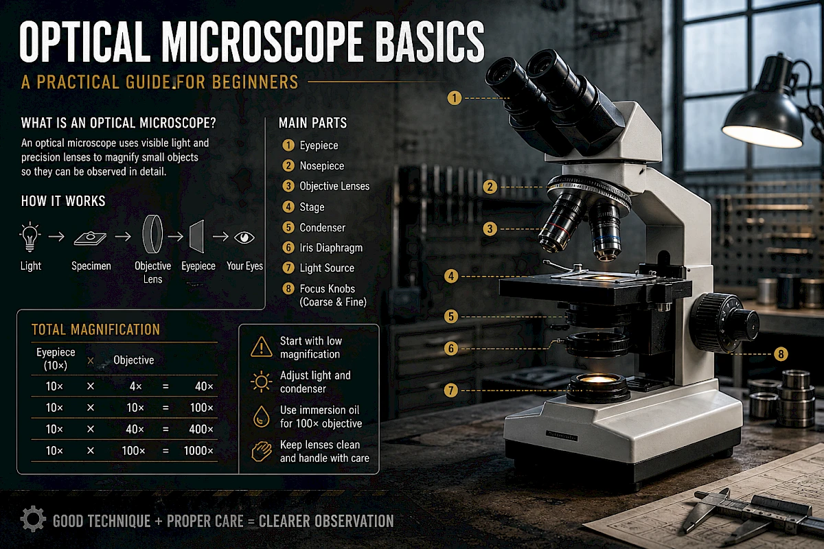
Light passes through or reflects from the specimen. The objective lens collects this light and forms a magnified image. The eyepiece then further magnifies that image for the observer.
In a standard compound microscope, total magnification is calculated as:
Total Magnification = Eyepiece Magnification × Objective Magnification
For example:
- 10× eyepiece × 4× objective = 40× total magnification
- 10× eyepiece × 10× objective = 100× total magnification
- 10× eyepiece × 40× objective = 400× total magnification
- 10× eyepiece × 100× objective = 1000× total magnification
However, higher magnification does not always mean better image quality. Resolution, contrast, illumination, lens quality, and sample preparation are equally important.
3. Main Parts of an Optical Microscope
Eyepiece
The eyepiece is the lens you look through. Most microscopes use 10× eyepieces. Some models may use 15× or 20× eyepieces, but stronger eyepieces do not automatically improve detail.
A good eyepiece provides a comfortable field of view and clear image edges.
Objective Lenses
Objective lenses are the most important optical components of the microscope. They are mounted on the rotating nosepiece and usually include:
- 4× scanning objective
- 10× low-power objective
- 40× high-power objective
- 100× oil immersion objective
The objective lens determines most of the microscope’s resolution, working distance, brightness, and image quality.
Nosepiece
The revolving nosepiece holds the objective lenses and allows users to switch magnification. It should click firmly into position. If the objective is not fully aligned, the image may appear dark, blurry, or partially blocked.
Stage
The stage supports the slide or sample. Most laboratory microscopes use a mechanical stage, allowing precise movement in the X and Y directions.
Smooth stage movement is important when scanning a slide or locating a specific area.
Condenser
The condenser focuses light onto the specimen. It plays a major role in brightness, contrast, and resolution.
Many beginners ignore the condenser, but incorrect condenser height can make even a good microscope perform poorly.
Iris Diaphragm
The iris diaphragm controls the amount and angle of light entering the condenser.
Opening it fully gives more brightness but may reduce contrast. Closing it too much increases contrast but can reduce resolution and create visual artifacts.
A well-adjusted diaphragm produces a balanced image with good detail and natural contrast.
Light Source
Modern microscopes usually use LED illumination. Older models may use halogen lamps.
LED lights are stable, energy-efficient, long-lasting, and generate less heat. Halogen lamps often provide warm color rendering but require more frequent replacement.
Coarse and Fine Focus Knobs
The coarse focus knob moves the stage quickly and is mainly used at low magnification.
The fine focus knob moves the stage slowly and precisely. It is especially important when using 40× or 100× objectives.
At high magnification, only the fine focus knob should be used to avoid damaging the slide or objective lens.
4. Common Types of Optical Microscopes
Compound Microscope
This is the most common type used in biology, education, and clinical laboratories. It uses transmitted light to observe thin, transparent, or stained specimens.
Best for:
- Cells
- Tissue sections
- Microorganisms
- Blood smears
- Prepared slides
Stereo Microscope
A stereo microscope provides a three-dimensional view at lower magnification. It is useful for observing larger objects that do not need to be sliced thinly.
Best for:
- Insects
- Plants
- Electronic components
- Small mechanical parts
- Jewelry
- Industrial inspection
Inverted Microscope
In an inverted microscope, the objectives are below the stage and the light source is above. It is commonly used for observing cells in culture dishes or flasks.
Best for:
- Cell culture
- Live cells
- Liquid samples
- Petri dishes
- Microplates
Metallurgical Microscope
This microscope uses reflected light to observe opaque materials.
Best for:
- Metals
- Ceramics
- Coatings
- Polished surfaces
- Failure analysis
- Semiconductor inspection
Fluorescence Microscope
A fluorescence microscope uses specific wavelengths of light to excite fluorescent dyes or naturally fluorescent structures.
Best for:
- Immunofluorescence
- Protein localization
- Cell imaging
- Microbiology
- Research applications
5. Magnification vs. Resolution
Many people believe that a microscope with higher magnification is always better. This is a common misunderstanding.
Magnification makes an image larger.
Resolution determines whether two close points can be distinguished as separate details.
If the microscope has poor resolution, increasing magnification only makes a blurry image larger. This is called empty magnification.
A microscope with good optics, proper illumination, correct condenser adjustment, and a well-prepared specimen can produce better results at 400× than a poorly adjusted microscope at 1000×.
6. What Is Numerical Aperture?
Numerical aperture, often written as NA, describes the light-gathering ability and resolving power of an objective lens.
A higher NA usually means:
- Better resolution
- Brighter image
- Shallower depth of field
- More demanding focus adjustment
For example, a 40× objective with NA 0.65 usually provides better detail than a 40× objective with NA 0.40.
When choosing objectives, magnification is not the only number that matters. Numerical aperture is just as important.
7. Dry Objectives and Oil Immersion Objectives
Most microscope objectives are dry objectives. They are used with air between the lens and the slide.
The 100× objective is often an oil immersion objective. It requires immersion oil between the objective lens and the cover glass.
Oil immersion improves resolution because immersion oil has a refractive index closer to glass than air does. This allows more light to enter the objective lens.
Important rules for oil immersion:
- Use only proper microscope immersion oil
- Apply only a small drop
- Never use oil with 4×, 10×, or 40× dry objectives unless they are specifically designed for oil
- Clean the 100× objective immediately after use
- Never let oil dry on the lens
Dried immersion oil is difficult to remove and may permanently affect image quality.
8. Brightfield Microscopy
Brightfield is the most common observation method. The background appears bright, and the specimen appears darker or colored.
It is suitable for stained samples, such as:
- Blood smears
- Tissue sections
- Bacteria stains
- Plant sections
Unstained transparent samples may show poor contrast under brightfield observation. In that case, phase contrast, darkfield, or staining may be needed.
9. Basic Steps for Using a Microscope
Step 1: Start with the Lowest Objective
Always begin with the 4× or 10× objective. Low magnification provides a wider field of view and makes it easier to locate the specimen.
Step 2: Place the Slide Correctly
Put the slide on the stage and secure it with the slide holder. Make sure the cover glass faces upward if using a standard upright microscope.
Step 3: Adjust the Light
Turn on the light and set it to a comfortable brightness. Avoid using maximum brightness unnecessarily.
Step 4: Focus Slowly
Use the coarse focus knob at low magnification. Once the image appears, use the fine focus knob to sharpen it.
Step 5: Adjust the Condenser and Diaphragm
Raise the condenser close to the slide for high-resolution observation. Adjust the iris diaphragm until the image has good contrast without losing detail.
Step 6: Increase Magnification
Rotate to the next objective. Most microscopes are parfocal, meaning the image should remain nearly focused when switching objectives. Only small fine-focus adjustments should be needed.
Step 7: Use Fine Focus at High Magnification
At 40× and 100× objectives, avoid using coarse focus. The working distance is very short, and rough movement may break the slide or damage the lens.
10. Common Beginner Mistakes
Using High Magnification Too Early
Starting with 40× or 100× makes it difficult to find the specimen. Always begin with low magnification.
Using Too Much Light
Excessive brightness can wash out details and cause eye fatigue. Proper contrast is more important than maximum brightness.
Ignoring the Condenser
A poorly adjusted condenser can make the image dim, uneven, or low in resolution.
Touching Lenses with Fingers
Fingerprints contain oil and moisture. They can reduce image clarity and damage lens coatings over time.
Using the Wrong Cleaning Material
Paper towels, tissues, clothing, or cotton swabs may scratch optical surfaces. Use proper lens paper or approved optical cleaning materials.
Forgetting to Clean Immersion Oil
Immersion oil left on the lens can harden and degrade optical performance.
11. What Makes a Good Microscope Image?
A good microscope image should have:
- Even illumination
- Sharp focus
- Suitable brightness
- Good contrast
- Minimal glare
- Natural color
- Clear field edges
- No dust spots or oil smears
If the image looks poor, do not immediately blame the microscope. Check the slide, focus, condenser, diaphragm, light intensity, objective cleanliness, and eyepiece cleanliness first.
12. Basic Troubleshooting
The Image Is Too Dark
Possible causes:
- Light intensity is too low
- Condenser is too low
- Iris diaphragm is too closed
- Objective is not clicked into position
- Dirty lens
- Incorrect filter or light path setting
The Image Is Blurry
Possible causes:
- Slide is upside down
- Objective lens is dirty
- Eyepiece is dirty
- Condenser is incorrectly adjusted
- Cover glass is too thick
- Wrong immersion oil technique
- Sample is too thick
There Are Spots in the Image
Possible causes:
- Dust on eyepiece
- Dust on camera sensor
- Dirt on objective lens
- Contaminated slide
- Air bubbles in immersion oil
A simple way to locate dust is to rotate the eyepiece. If the spot rotates, it is on the eyepiece. If it stays still, check the objective, slide, or camera path.
The Image Has Poor Contrast
Possible causes:
- Iris diaphragm too open
- Sample is unstained
- Light too bright
- Condenser not adjusted
- Wrong observation method
13. Choosing the Right Microscope
Before buying or selecting a microscope, consider the application first.
For biology teaching, a standard compound microscope with 4×, 10×, 40×, and 100× objectives is usually enough.
For cell culture, an inverted microscope is more suitable.
For electronic parts, insects, or solid objects, a stereo microscope is the better choice.
For metal surfaces, a metallurgical microscope is needed.
For fluorescence labeling, a fluorescence microscope is required.
A microscope should be chosen based on the sample, not only on magnification.
14. Practical Tips for Better Observation
- Start with low magnification
- Keep lenses clean
- Adjust illumination carefully
- Use the correct condenser height
- Do not over-close the diaphragm
- Use proper slides and cover glasses
- Avoid thick or uneven samples
- Focus slowly at high magnification
- Clean immersion oil immediately after use
- Cover the microscope after work
Good microscopy depends on both equipment quality and user technique.
Conclusion
An optical microscope is not just a magnifying device. It is a precision optical instrument that requires correct setup, careful focusing, proper illumination, and regular care.
For beginners, the most important habits are simple: start with low magnification, adjust the light properly, keep lenses clean, and handle the microscope gently.
Once these basics are understood, microscope observation becomes easier, more accurate, and much more reliable.